Life Sciences
Cell Culture & Assay
Our cultureware products enable researchers to create and analyze unique and informative experimental models.
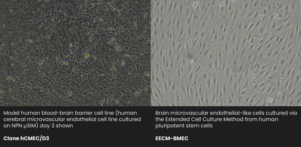
Barrier Cells
Live Cell Culture Imaging
- Observe live cell dynamics with unparalleled clarity: Watch cells move over or through porous membranes with outstanding contrast, including immune cell transmigration across endothelial and epithelial barriers.
- Easily monitor cell growth: Follow the growth of cells to confluency right on the membrane using conventional microscopy.
Time-lapse phase contrast images were recorded of neutrophils migrating through a nanoporous membrane-supported HUVEC monolayer. Phase-bright neutrophils are on the Lumenal side, while phase-dark neutrophils have translocated to the ablumenal side of the HUVEC layer.
Image Credit: https://www.ncbi.nlm.nih.gov/pmc/articles/pmid/30632319/
Immunofluorescence Studies
- Label and image without having to cut out membranes: Unlike traditional membrane inserts, directly immunolabel one or more cell types, on either membrane surface, and image without any additional bothersome manipulations.
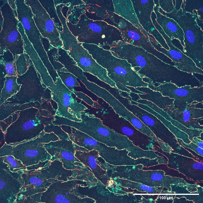
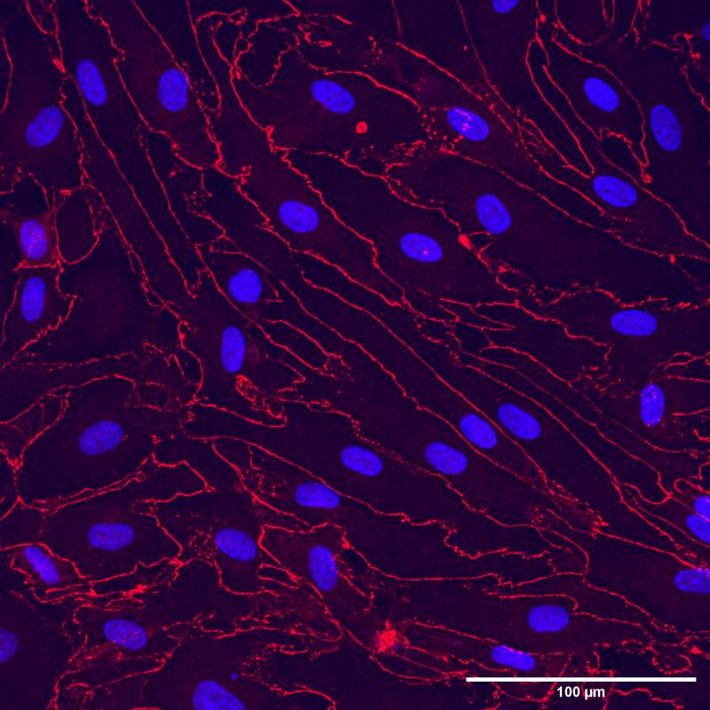
IPSC-derived endothelial-like cells cultured on Silicon Nanomembranes were fixed and stained for tight junction proteins (Claudin-5 and ZO-1), and counterstained with Hoechst 33342. Cells were imaged with a confocal microscope at 40X magnification.
Image Credit: Molly McCloskey and James McGrath, University of Rochester
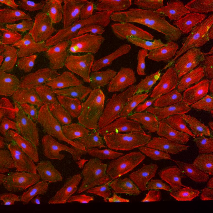
Confocal Microscopy of Co-Cultures
- See cells and basement membranes, not plastic: Nanoporous silicon nitride membranes are thinner than an optical slice in confocal microscopy, so are nearly invisible in confocal reconstructions of co-cultures and basement membranes.
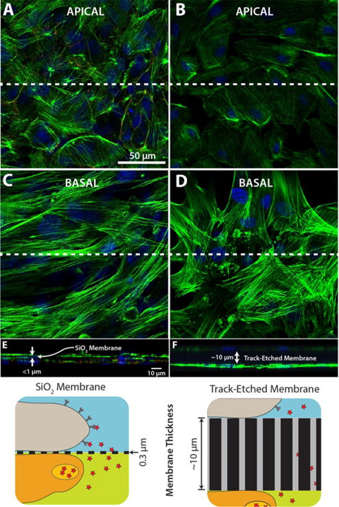
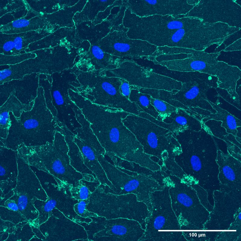
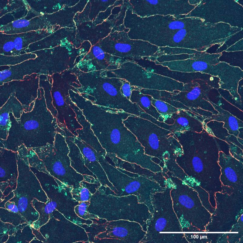
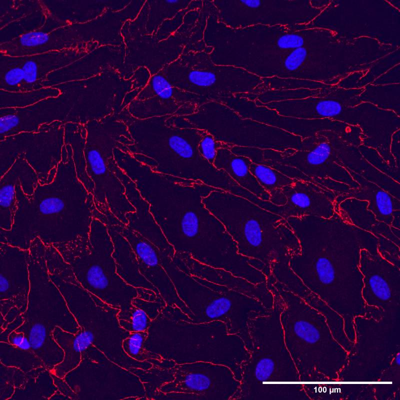
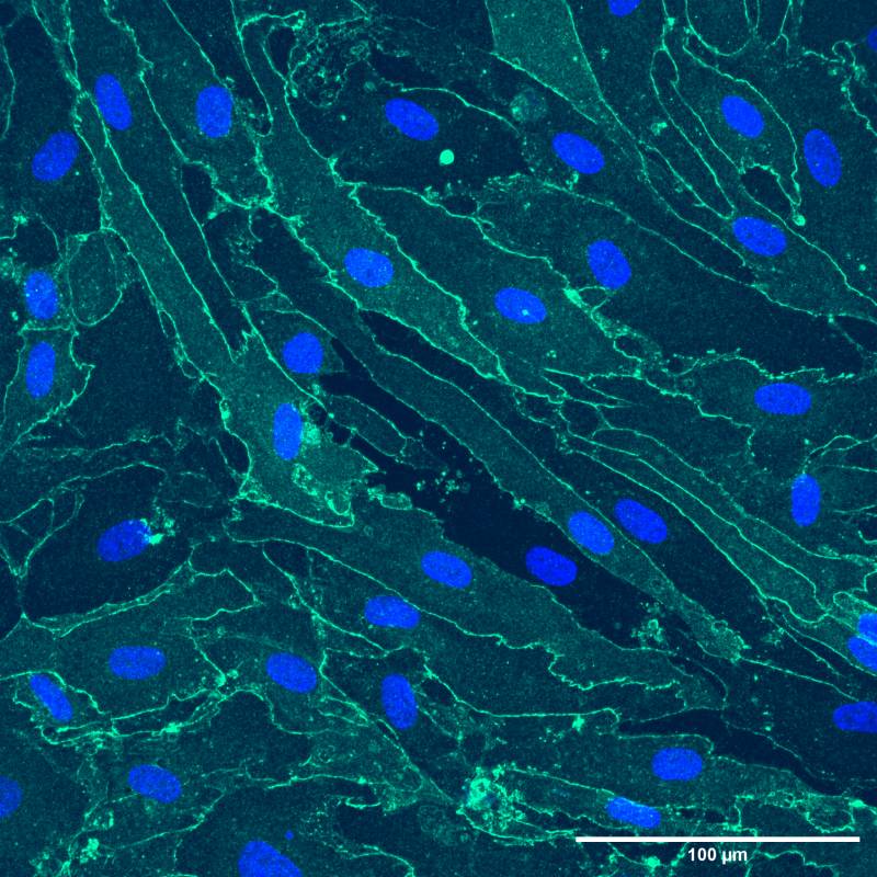
Fluorescent images of endothelial (HUVEC) and adipose stem cells (ADSCs) grown on silicon nanomembranes. Cells were fixed, permeabilized, and stained for nuclei (blue), actin cytoskeleton (green), and VE-cadherin (red). (A) ADSC grown on the apical side and (C) HUVEC grown on the basal side of a silicon nanomembrane. (B) ADSC grown on the apical side and (D) HUVEC grown on the basal side of a conventional membrane insert. (E, F) Cross-sections taken at the dashed white line.
Image Credit: https://www.ncbi.nlm.nih.gov/pmc/articles/PMC5373106/
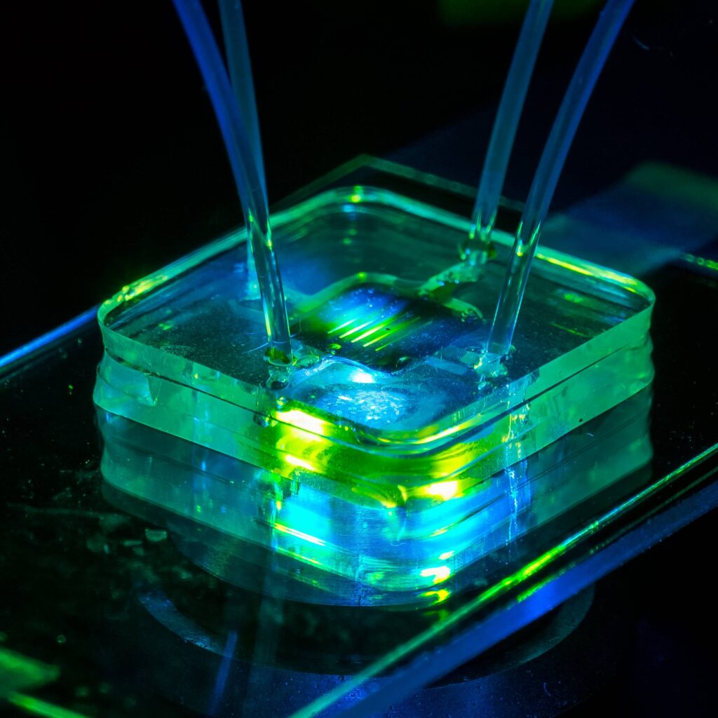
Compartmentalized Cultures
- Paracrine communication as if there were no membrane: Extraordinarily permeable silicon nanomembranes support small molecule diffusion between apical and basal compartments without any measurable hindrance from the membranes.
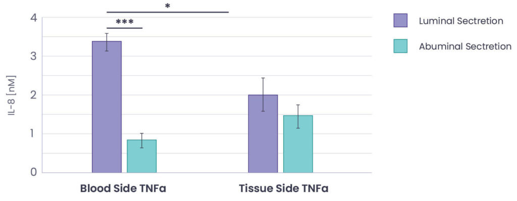
An in vitro vasculature model was created on a silicon nanomembrane, permitting stimulation by an inflammatory molecule from either the top (lumenal) or bottom (ablumenal) chambers. The media was harvested from either chamber and a standard ELISA was used to measure IL-8 secreted into either chamber.
Image Credit: https://doi.org/10.3389/fmedt.2020.600616
Single Cells, Spheroids & Organoids
Microbubble Array™ products promote a wide range of research activities due to their unique features.
- One-of-a-kind hemispherical microcavities within a cell-compatible substrate
- Cavities with nanoliter volumes are ideal for creating microenvironments in which to culture and assay single-cell and multicellular spheroids and organoids
- Useful for stem cell and antibody-producing cell clonal selections
- Available in convenient Chamber Slide, Multi-Well plate, and Culture Dish formats with various cavity opening and volume sizes
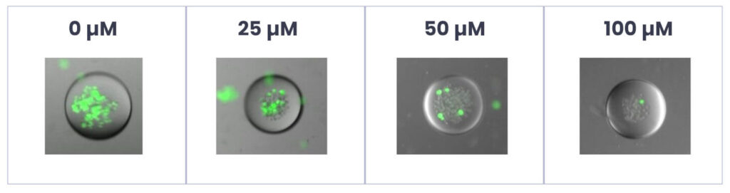
MB arrays can be used to identify drug-resistant cancer cells
MC-1 is a melanoma cancer cell line derived from lung metastasis. MC-1s grown in a hydrophobic environment displayed a nonadherent phenotype. Images of MC-1 melanoma cells cultures after 48 hours in increasing Cisplatin treatment. Live cells are stained with green Calcein-AM.
Cultureware Options
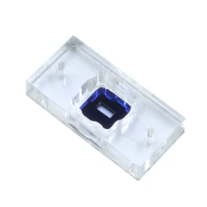
Get the highest resolution imaging of barrier-forming cells with SiMPore’s µSiM-CellVu products.
µSIM-CellVu Options of Our Microfluidics Enabled by Silicon Membranes Products
µSiM-CellVu Cultureware is available in three formats, enabling shorter working distance and higher magnification imaging:
| Format | Working Distance | Magnification |
|---|---|---|
| Standard | 600 µm | Up to 40X LWD |
| High-Resolution | 300 µm | Up to 40X |
| Ultrahigh-Resolution | 225 µm | Up to 100X |
We also offer a choice of membrane pore sizes for experimental flexibility:
| Membrane | Pore Size | Recommended Use |
|---|---|---|
| Nanoporous | 50 nm | Barrier cell models |
| Dual-Scale | 50 nm + 3 µm | Transmigration studies |
| Microporous | 500 nm, 3 µm | Miscellaneous cell assays |
Shop µSiM-CellVu Products
Purchase a complete µSiM-CellVu assembly, build your own with a variety of µSiM kit options, or shop individual µSiM components.