
Authored by: Teagan Horan, Applications Scientist, SiMPore and Peng Miao, Applications Scientist, HORIBA Instruments Inc.
Introduction
USP788 is an accepted method by which to test parenteral drug products for the presence of subvisible particles 10 µm and larger. Method I utilizes light obscuration to detect and measure levels of these particles. When samples exceed the acceptable threshold level for subvisible particles (thus failing Method I’s criteria), practitioners must then perform forensic investigations utilizing sample filtration and microscopic analysis techniques outlined in Method II. This present study examined the utility of silicon nanomembranes and Raman spectroscopy to carry out forensic Method II investigations, and further examined the benefits of multi-modal analyses enabled by silicon nanomembranes for such investigations.
Particle Identification Efficacy
SiMPore’s 1 µm cut-off silicon nanomembranes in 25 mm filter disks and 25 mm black-dyed PCTE filters (currently recommended for use with Method II) were each dosed in triplicate with a known concentration of 10-30 µm ETFE particles (NIST reference standard #8634). Filters were imaged in 10x darkfield and particles counted via HORIBA’s ParticleFinderTM software in LabSpec6. Utilized active areas of both filters were made equivalent by cutting custom PDMS gaskets so that particle counts on both substrates would be comparable.
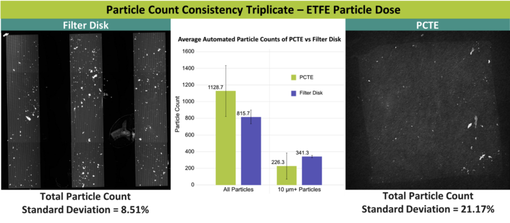
Higher consistency particle counts were readily observed on silicon nanomembranes than on PCTE membranes, with a 40% improvement in the counts’ standard deviation.
During the PCTE particle counts, multiple software settings had to be adjusted, including the threshold slider, the threshold mathematical model base, and several particle morphological prefilters. Such settings need to be adjusted for all technical replicates of the PCTE dataset in order to distinguish pores from particles and reduce item misidentifications.
Silicon nanomembranes only required adjustment to the threshold slider, as technical replicates were very similar in terms of particle vs. pore threshold values. For PCTE, there are likely pore and particle misclassifications in the data, which is reflected in the wider count standard deviation, as compared to counts on silicon nanomembranes.
Forensic USP788 Method II Mock Sample Study
We generated a set of surrogate solutions replicating a pharmaceutical product that would pass or fail USP788 Method II. This sample consisted of dissolving bovine serum albumin (BSA) into ultrapure water, then vortexing the suspension to cause aggregation. Additional samples were also thermally stressed to create additional aggregates in size and count.
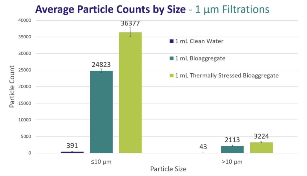
Three 1 mL aliquots of the BSA aggregate suspension were filtered onto 1 µm cut-off, gold-coated silicon nanomembranes within 25 mm filter disks. Captured particles were counted via HORIBA’s LabSpec6 ParticleFinderTM software. As shown above, consistent particle counts were observed, with variation increasing as particle count neared 40,000 for the sub-10µm particles in the thermally stressed sample. Only the threshold slider required adjustment to identify particles. Based on this initial result, gold-coated silicon nanomembranes appear to be a suitable substrate for forensic particle composition investigations.
Multimodal Forensic Analyses
Production lots that fail either USP788 Method may require more extensive particle analysis by multiple modalities in order to find the root cause of such production failures. Raman spectroscopy and scanning electron microscopy (SEM) may elucidate the exact means by which contaminants formed or entered into the product, enabling faster and more efficient solution implementation.
We generated a sample to mimic a failed pharmaceutical product containing intrinsic and extrinsic contaminant types for both visual and spectroscopic identification.
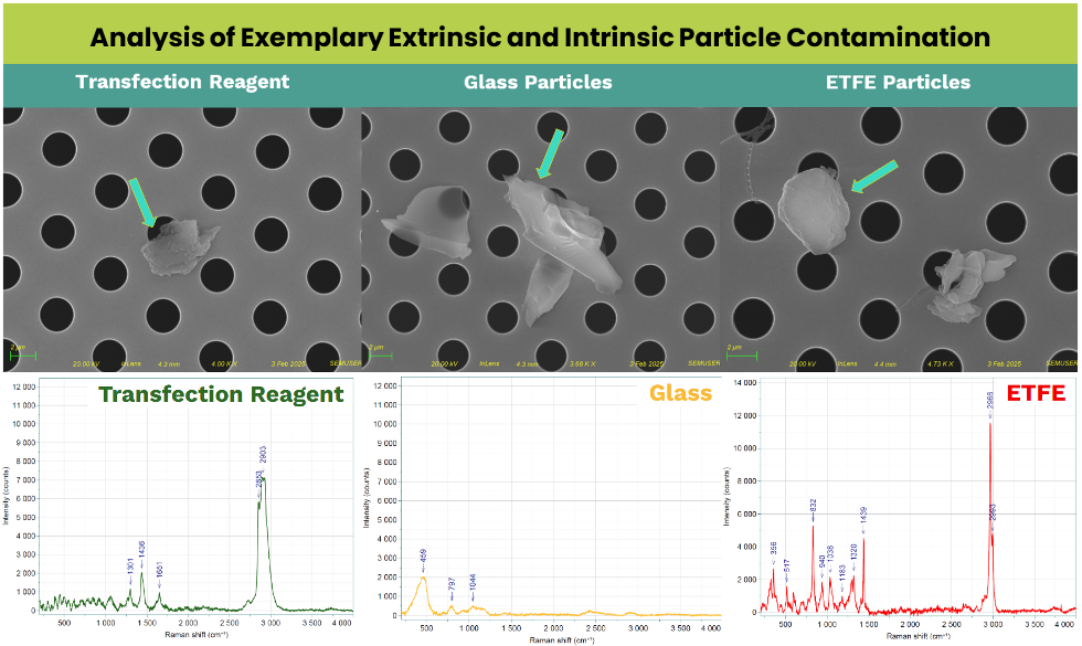
For these purposes, “Intrinsic particles” refer to particles that are generated wholly from within the sample (e.g., aggregates), while “extrinsic particles” refer to contamination that enters from outside (e.g., glass shards from packaging vials; fibers shed by process filters). Cationic lipids (typically used in transfection reagents) suspended in ultrapure water were thermally stressed and vortexed to create lipid aggregates to mimic intrinsic particles. Additionally, ETFE reference particles (NIST #8634) and <10 µm glass shards were spiked into the lipid aggregate suspension to mimic intrinsic and/or extrinsic contaminants (depending on product type and final packaging).
The lipid aggregate + contaminants suspensions were filtered onto 3.0 µm cut-off, gold-coated silicon nanomembranes within 25 mm filter disks, then the captured particles were analyzed with SEM and Raman spectroscopy. By localizing the particles with coordinate-based mapping, the same particles could be analyzed by either modality. Raman spectroscopy was able to identify particle composition, while SEM confirmed morphology of the captured particles – all of which reveal clues to particle sources.
Conclusions
Method I of USP788 is unable to provide root cause information when production lots exceed the allowable particle threshold level specified under Method I, as light obscuration cannot provide critical particle composition and identity information. Conventional PCTE substrates recommended for routine Method II measurements are also unable to support the extensive particle characterizations needed to determine particle composition and/or morphology, so that problems can be readily identified and mitigated as expeditiously as possible. Here, we present a workflow enabled by silicon nanomembranes, Raman spectroscopy, and SEM analytical modalities that empower practitioners with the resources for tackling USP788 root cause failure analyses.
Methods
- 800 µm thick clear-cast PDMS was cut to user-defined size to match desired surface area allowances.
- Figure 1 Microscopy imaging conducted on an Olympus BX61 and image processing done through ImageJ.
- Particle analysis conducted utilizing ParticleFinder via HORIBA’s LabSpec6.
- Raman analysis conducted utilizing HORIBA’s XploRA plus instrument enabled with LabSpec6.
- Pharmaceutical analogues were generated by mixing 1.5 g of BSA into 30 mL of 0.22 µm filtered MilliQ water and vortexing for 30s to mix. Thermal stress induced via 60°C overnight incubation. Both samples were chilled at 4° for 48 hrs. Sample then filtered on 1 µm gold coated SiMPore silicon nanomembranes.
- Additional intrinsic particle samples generated by thermally and physically stressed Polysorbate-80. Thermal stress was induced via 3 day incubation at 60°C and once a day vortex for 1 minute during incubation.
- Graphs are showcased in terms of the major axis of each identified particle for each sample. The average 3 trials are shown with error bars denoting standard deviation of the triplicates.
- SEM micrographs taken utilizing a Zeiss Auriga system.
Acknowledgements
This study was funded in part with support from the National Institute of Environmental Health Sciences of the National Institutes of Health under Award Number R44ES031036.
The content is solely the responsibility of SiMPore Inc. and does not necessarily represent the official views of the National Institutes of Health.
Data was collected with help from HORIBA.
About SiMPore
SiMPore’s mission is to develop and deliver products for the analysis of nanoscale specimens, enabling the discovery of the future in material and life sciences. The Company offers a variety of filters for capture and analysis of particulates of interest, as well as substrates for electron microscopy.
About HORIBA
HORIBA develops unique measurement and analysis technologies to support a wide range of industries and applications that address global societal challenges. As leaders in Raman technologies, HORIBA delivers comprehensive solutions that extend beyond measurement and analysis across three megatrend-driven business fields: Energy & Environment, Bio & Healthcare, and Materials & Semiconductor.